
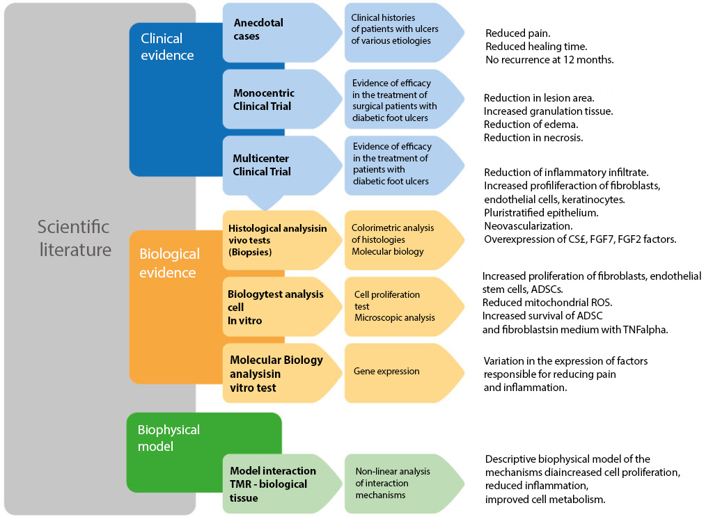
TMR® Scientific Validation.
THERESON products are designed for home use that can ensure patient compliance with therapeutic treatment and ease of use, in the comfort of their own home. For the following reasons, we have provided the customized rental formula that best suits the needs of our patients.
The formula provides for device rental starting from one month, which can be adjusted to the patient’s course of treatment. In addition, in case of prolonged use, the option of purchase is proposed. In the case of purchase after the first rental phase, a discount will be applied that will take into account the period of rental made.
To request a free quote use the form below.
Single-center study of surgical patients
The study was conducted by Prof. A. Piaggesi at the University of Pisa (Italy) and showed statistically significant evidence of acceleration in wound healing (1). The results were published by a leading scientific journal called “ The International Journal of Lower Extremity Wounds ” at the end of March 2015.
Methods
Two groups of 10 patients, admitted for surgery, were selected to check some clinical and biological parameters that changed as a result of TMR therapy:
- Both groups of type 2 diabetic patients with post-surgical lesions with similar area greater than 1 cm2, located distal to the ankle and with a low level of ischemia.
Both groups received standard treatment (initial surgical debridement, revascularization if necessary, and Hydrofiber bandaging).
One group received additional treatment with TMR® for two consecutive weeks.
Results
The reported results showed an increased a healing rate, a reduction in wound surface area, and a reduction in pain, as can be seen in the following graphs extracted from the published study.
The healing parameters that improved with TMR therapy were:
- Healing Rate at 6 months was 90% in the TMR®-treated group and 30% in the group treated only with single standard treatment (statistically significant; p < 0.05).
Healing time in the TMR®-treated group was 84.46 days (with a standard deviation of 54.38) versus 148.54 days (with a standard deviation of 78.96) for the control group (statistically significant; p < 0.01).
At the end of a 12-month follow-up period, all patients in the treated group were healed, while three patients in the control group were still ulcerated.
Pain measured by VAS scale was significantly (p < 0.05) reduced faster in the TMR®-treated group than in the group that received standard treatment alone, in the first 3 months of follow-up.
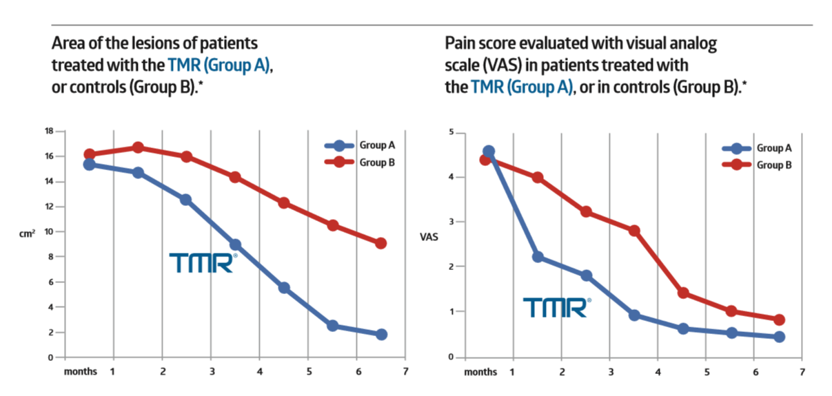
The statistically significant results of this clinical study observed that TMR® treatment supplementary to standard care achieves healing faster than standard therapy alone.
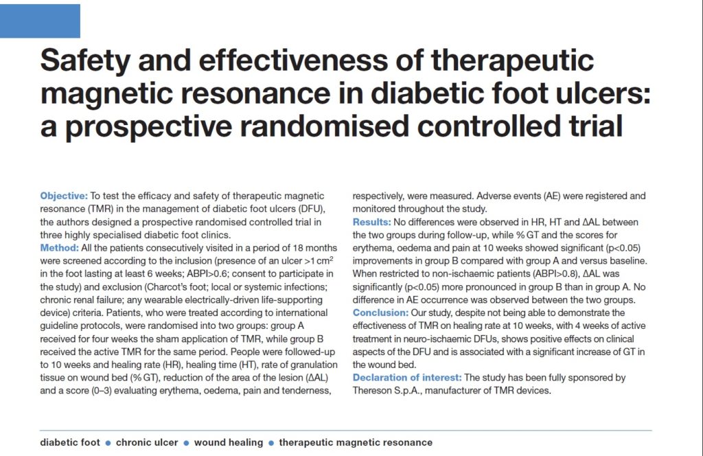
Multicenter exploratory clinical study
Clinical experience in using TMR® was proven in a double-blind exploratory clinical study, in which a dramatic reduction in patients’ lesion areas and volumes (compared with standard treatment alone) was shown.
A total of 157 patients were enrolled in 3 centers:
- Pisa (Cisanello – Prof. A. Piaggesi),
Treviso (Ca’ Foncello Hospital – Dr. M.Sambataro),
Peschiera del Garda VR (Casa di Cura Pederzoli – Dr. C.Nicoletti).
In a moderately ischemic subgroup, the markedly greater reduction in lesion area (75% vs. 19% in the control group) with sham device, reduction in pain (VAS scale), edema, necrotic tissue and increase in granulation tissue is shown.
The results of these studies were published in December 2016 in the Journal of Wound Care (JWC) under the title: “ Safety and Effectiveness of Therapeutic Magnetic Resonance in the Management of Postsurgical Lesion of the Diabetic Foot.” (2)
In vivo Biopsy Study
Tests performed on biopsies showed that treatment with TMR® produced a statistically significant increase in proliferative cells such as keratinocytes, endothelial cells and fibroblasts, in treated patients, acting simultaneously in reducing inflammation and thus the inflammatory infiltrate.
After 15 days of TMR® treatment, histo-pathological analyses showed tissue reorganization and differentiation with the formation of dermal papillae and new blood vessels.
The results of these studies were submitted for review by the journal “Regenerative Medicine” in June 2015. The provissory title of the article is: “Histological and molecular analyses of granulation tissue of diabetic foot ulcers treated with TMR ”.
Gene expression results of growth factors relevant to cell proliferation and wound healing were also included in the article. Indeed, some factors showed significant overexpression in TMR®-treated patients compared with untreated patients.
(1) Source: Lorenza Abbruzzese, DPM, Elisabetta Iacopi, MD, Alberto Coppelli, MD, Giovanni Bonino, DPM, Chiara Goretti, MD, and Alberto Piaggesi, MD, Safety and Effectiveness of Therapeutic Magnetic Resonance in the Management of Postsurgical Lesion of the Diabetic Foot, The International Journal of Lower Extremity Wounds, March 2015,14: 4-10.
(2) Fonte: A. Piaggesi, MD; M. Sambataro,MD; C Nicoletti, MD; C. Goretti, MD; E. Lacopi, MD; A. Coppelli, MD, Safety and effectiveness of therapeutic magnetic resonance in diabetic foot ulcers: a prospective randomised controlled trial, Journal of Wound Care, Vol 25 , No 12 , December 2016.
University of Padua – Italy.
Prof. Rizzuto, Prof. Zavan Department of Biomedical Sciences
In vitro tests.
In vitro tests in healthy and diabetic tissues showed increased proliferation of fibroblasts and endothelial cells after exposure to TMR® treatment.
Preliminary results showed that the TMR method, through daily treatments, promotes increased proliferation of fibroblasts and endothelial cells, the type of cells that are involved in wound healing processes.
These results were published online on June 5, 2015 by the scientific journal “Journal of Tissue Engineering and Regenerative Medicine” with IF 5,199. The scientific article with all the results is titled “Treatment by Therapeutic Magnetic Resonance increases fibroblastic activity and keratinocyte differentiation in an in vitro model of 3D artificial skin.”
As an additional positive sign, exposure to TMR® also showed a reduction in oxidative stress condition in both fibroblasts and endothelial cells (3).
Following some indications from patients using the device and the abundant literature on the subject, tests were performed to verify the efficacy of TMR® electromagnetic fields in bone cells.
The study containing the results of these tests shows that TMR® is able to induce increased proliferation of osteocytes and overexpression of key osteogenic genes, particularly osteocalcin, osteonectin, ostepontin and alkaline phosphatase. TMR® also promotes the deposition of hydroxyapatite crystals in bone matrix. This study was published in March 2016 in the journal Life sciences under the title, “Pulsed magnetic therapy increases osteogenic differentiation of mesenchymal stem cells only if they are pre-committed.” (4)
(3) Source: Ferroni L, Bellin G, Emer V, Rizzuto R, Isola M, Gardin C, Zavan B, Treatment by Therapeutic Magnetic Resonance (TMR™) increases fibroblastic activity and keratinocyte differentiation in an in vitro model of 3D artificial skin, J Tissue Eng Regen Med. 2015 Jun 5. doi: 10.1002/term.203
(4) Source: Letizia Ferroni, Ilaria Tocco, Andrea De Pieri, Martina Menarin, Enrico Fermi, Adriano Piattelli, Chiara Gardin, Barbara Zavan. Pulsed magnetic therapy increases osteogenic differentiation of mesenchymal stem cells only if they are pre-committed, Life Sciences 152 (2016) 44-51
Bogolyubov Institute of Theoretical Physics, Kiev – Ukraine.
Prof. Brizhik.
Identification of mechanisms of interaction between pulsating magnetic fields (PEMFs) and redox processes in body tissues.
Prof. Brizhik is collaborating with the University of Padua, Department of Biomedical Sciences, to explain how TMR® therapy with low-intensity magnetic fields, oscillating well-defined frequencies, is able to influence biological processes such as soft tissue healing.
A literature review conducted by Prof. Larissa Brizhik on the ‘interaction of PEMFs with biological tissues was published on November 26, 2014, in the Journal of Advances in Physics (5).
An article (6) describing the biophysical working principle of TMR® therapy was also published in the “International Journal of Biophysics.” The article clarifies how from clinical evidence it was possible to formulate a theory involving the influence of electromagnetic fields at the cellular level via a form of energy transport discovered in 1834 by Scott Russel: the soliton. The approach developed by Prof. Brizhik and Eng. E. Fermi is supported by the various ongoing biological and clinical studies. The title of the article is “On the Mechanisms of Wound Healing by Magnetic Therapy: The Working Principle of Therapeutic Magnetic Resonance.”
(5) Fonte: L. Brizhik, Effects of magnetic fields on soliton mediated charge transport in biological systems, Journal of advances in Physics, Vol.6, No.2, 26 November 2014, 1191-1201.
(6) Fonte: Larissa Brizhik, Letizia Ferroni, Chiara Gardin, Enrico Fermi, On the Mechanisms of Wound Healing by Magnetic Therapy: The Working Principle of Therapeutic Magnetic Resonance, International Journal of Biophysics 2016, 6(3): 27-43

Pursuant to the Ministry of Health Circular dated 28/03/2013, THERESON S.p.A. warns the user that the content on the site (text, images, photos, etc.) is aimed exclusively at health professionals and has an exclusively informative content. Therefore, such information does not qualify as advertising.
THERESON S.p.A. disclaims any responsibility for consultations by non-professionals.
Request information
Our technicians, salesmen and doctors are at your disposal for any clarification on this matter. Please fill out the form or contact us directly.